
Adult Eye Exams
ADULT EYE EXAMS
Why eye exams are so important
A comprehensive adult eye exam allows your eye doctor to assess your vision, ensure any prescription lenses you’re wearing are accurate and make sure your eyes are healthy.
Since many common eye diseases develop asymptomatically, your eye doctor can detect and manage any issues at an earlier stage, thereby giving you the best chance of protecting you against vision loss.
We recommend our adult and senior patients be examined every 1–2 years, but if you have a very high or fluctuating prescription, a diagnosed eye disease, or other risk factors that may affect your visual health, we will recommend a personalized schedule.

What to expect at your eye exam
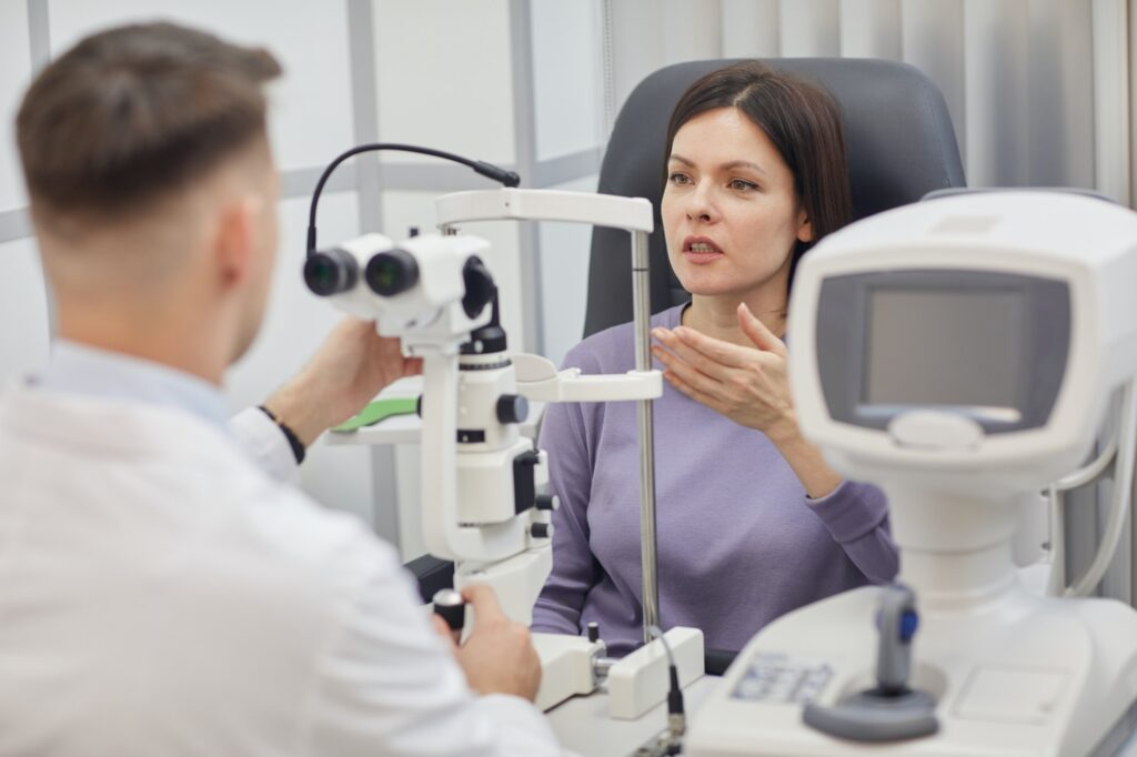
- Our trained technician will begin by collecting preliminary eye measurements utilizing the latest technology to make your examination as accurate as possible. These include measurement of any potential refractive error, like nearsightedness and astigmatism, then testing of the pressure inside your eyes that could indicate glaucoma. Next, optional, but recommended, Optomap retinal imaging will be performed. This amazing technology allows your doctor to view almost the entire back of your eye in one single high-resolution image, and does not require your eyes to be dilated with eye drops. These images will be reviewed in the exam room with your doctor.
- Your eye doctor will begin the exam by getting to know you and your specific concerns, then review your history as well as the data collected by the technician.
- Your doctor will measure how clearly you are seeing, then will assess how well your eyes are working together as a team to be sure you have clear, comfortable vision throughout the day.
- Next, your doctor will complete a thorough health evaluation, including assessment of the front of your eye, looking for dry eyes, allergy, cornea disease and cataracts. Next, a thorough evaluation of the back of your eye will be done to be sure there is no disease, such as glaucoma and macular degeneration, amongst many other conditions.
- At the conclusion of the examination, your doctor will summarize their findings and recommendations and answer any questions you have.